
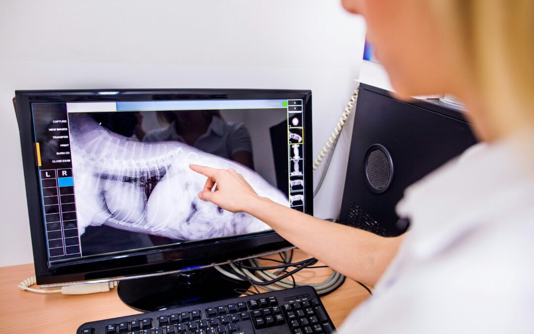
The trouble with our four-legged patients is that they can’t point to where it hurts. A limping dog might have a torn ligament, a small fracture, or arthritis. A vomiting cat could be dealing with anything from a hairball to a swallowed string causing serious damage. Diagnostic imaging at Douglas Animal Hospital lets us look past the symptoms and see exactly what’s going on inside, which means we can treat the right problem the first time around.
More Than Just X-Rays
When most pet owners think of imaging, they picture the classic chest x-ray. Radiographs are still one of our most useful tools, but veterinary diagnostics have expanded well beyond a single black-and-white film.
Digital radiography captures sharper images in seconds, with adjustable contrast that brings out details older film couldn’t show. Ultrasound uses sound waves to give us a live, moving view of soft tissue: the heart pumping, the bladder filling, a uterus being checked for puppies. Some cases call for CT or MRI scans through a specialist referral, particularly for complex orthopedic injuries or suspected brain conditions.
Each tool has a job. The right question isn’t “which is best” but “which one answers what we’re asking?”
What Imaging Actually Catches
A handful of the most common reasons we reach for imaging:
- Lameness or limping that doesn’t resolve. X-rays show fractures, hip dysplasia, elbow problems, and degenerative joint disease.
- Suspected foreign body ingestion. A rock, a hair tie, a corn cob piece, or a sock can show up on radiographs or appear as a telltale obstruction pattern in the gut.
- Persistent coughing or labored breathing. Chest x-rays reveal pneumonia, heart enlargement, fluid in the lungs, or masses.
- Repeated vomiting or weight loss. Abdominal imaging looks at organ size, shape, and structure, often catching kidney stones, tumors, or pancreatitis.
- Pregnancy confirmation and counting. Ultrasound confirms pregnancy early; late-term x-rays count puppies or kittens so you know what to expect during delivery.
- Dental problems below the gumline. Dental radiographs are the only way to see root abscesses, fractured tooth roots, or jaw bone loss in pets.
A physical exam tells us where the problem might be. Imaging tells us what the problem actually is.
When Your Vet Recommends Imaging
A recommendation for x-rays or ultrasound isn’t a sign of bad news. It’s often the fastest path to a real answer, which usually saves money compared to running multiple visits and treating symptoms blindly.
Take a senior dog with a sudden cough. Without imaging, we’d be guessing between several possibilities and trying medications one at a time. A quick chest film shows whether the heart is enlarged, the lungs are clear, or there’s a mass that needs further workup. That single image can shape weeks of treatment in a few minutes.
The same goes for a cat that stops eating. Bloodwork tells us how the organs are functioning at a chemical level. Ultrasound shows us what those organs look like physically, which is how we catch things like intestinal thickening from inflammatory bowel disease or the early shadows of cancer.
How Imaging at Douglas Animal Hospital Keeps Pets Safe
Most x-rays at our hospital are taken quickly, often without sedation, especially for cooperative patients. Animals that are anxious, painful, or need very precise positioning (such as hip x-rays for OFA certification) may need a mild sedative so we get a clear image without stressing them. Light sedation is also gentler on a pet’s joints when they’re already sore.
Modern veterinary radiography uses very low doses of radiation. Our team wears protective equipment and follows safety standards from the American College of Veterinary Radiology to protect both pets and staff.
Ultrasound is completely radiation-free. We shave a small patch of fur on the belly to get a clear picture, apply warm gel, and most pets settle in with a technician gently holding them. The whole exam usually takes 20 to 40 minutes depending on what we’re looking at.
What Happens After the Images
Once we have the pictures, the real value comes from reading them well. Our veterinarians review every study, and complex cases get a second look from a board-certified radiologist who reads the films remotely and sends a detailed report, usually within a day. That second set of eyes catches subtleties a generalist might miss and adds weight to the treatment plan we build for your pet.
If something concerning shows up, we go over the findings with you in plain language. You’ll see the images, understand what they mean, and know the options before any decisions get made.
Getting a Clearer Picture for Your Pet
Diagnostic imaging takes the guesswork out of veterinary medicine. A few minutes of a calm exam, a quick image, and we move from “something is wrong” to “here’s exactly what we’re dealing with.” That clarity leads to faster treatment, better outcomes, and far less worry for you.
If your pet is showing symptoms that don’t have an obvious explanation, or you’d like to talk through preventive screening for a senior pet, the team at Douglas Animal Hospital is happy to help you decide whether imaging makes sense for their situation. We’re here to help you understand what’s happening inside your companion, big or small, so they can get back to feeling like themselves.